
Imaging
Imaging at Barkat Cancer Hospital includes radiology, MRI, city simulator, etc
.The imaging department of Barkat Cancer Hospital is located on the negative first floor and includes MRI, radiology, CT scan, ultrasound, CT simulator, mammography and fluoroscopy departments
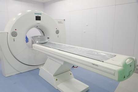
CT Scan
The name of this device is SIEMENS 128 SLAISE, and it is one of the newest devices available in the medical field, which provides the highest resolution and lowest noise image with the highest speed of bed movement and image processing in a fraction of a second. Among the capabilities of the device is the possibility of taking pictures of all the vessels of the body, especially the coronary arteries of the heart. According to the needs of the Barkat Comprehensive Cancer Center, which has made the diagnosis and treatment of cancer the criteria, this device provides the best image according to the specific conditions of the patient and the needs of the attending
physician
MRI
The name of this device is Tesla 1.5GE. Also, this device is the newest and only device available in Iran, with the ability to adapt to the conditions of patients undergoing radiation therapy. Because the imaging of such people, due to the sensitivity and precision of radiation therapy, requires special and unique arrangements and equipment, which this device fulfills. Among other exceptional features of this device, it is possible to point out that it has
the latest receiver and transmitter of electromagnetic waves (coil), which increases the quality of the image and comfort of the patient during imaging
Mammography
X-ray (radiography) is a non-invasive medical test that helps doctors diagnose and treat disease. In x-ray imaging, a part of the body is exposed to a small dose of ionizing radiation to produce pictures of the inside of the body. . Mammography is a specialized medical imaging that uses a low-dose x-ray system to view the inside of the breasts. Mammography test known as mammogram helps women in early diagnosis of breast diseases
Fluoroscopy device
Fluoroscopy is another imaging technique that is widely used in the diagnostic sciences of radiology and medical physics. The working basis of a fluoroscope device is based on an X-ray generator whose rays are received and amplified by a type of receiver called an image intensifier. This device detects X-rays passing through the patient's body in real time and displays and records them through a closed circuit television system.
It is also possible to display the image directly and in real time on a screen at times when movement in the body is desired (for example, checking the movement of contrast material in the gastrointestinal tract).